Vladimir Nesterov
Full member of Academy of Medical and Technical Sciences, Academician,
President of International Academy of non-linear diagnostic systems.
Primary osteoarthritis deformans of the knee-joint is one of the most pertinent problems in modern medicine due to its prelevance, great loss of working time and treatment expenses. In addition, in many cases an early or differential diagnosis of the knee-joint lesion is impeded, which complicates selecting the most efficient therapeutical and rehabilitation measures and evaluating the patient’s disability.
Modern diagnostics of knee-joint disorders comprises conventional radiography as well as sonographic evaluation of the joints, used to examine soft tissues of the locomotorium. The existing techniques used to examine the knee-joint allow to determine dominance of pathological process in the joint, including degenerative ones. However, the relationship between the intensity of pathomorphological changes and the severity and dynamics of the process have not been studied yet.
This paper aims to demonstrate the efficiency of the NLS-investigation in diagnosing osteoarthritis deformans, especially in the early (subclinical) phase of the disease.
To define a normal relationship of the knee-joint anatomical structures 10 healthy persons aged from 25 to 55 (test group) were examined. The main group consisted of 50 patients with clinical implications of osteoarthritis deformans of knee joints in different phases. The average duration of the disease was 7.0±3.0 years. All patients were routinely radiologically examined in two interperpendicular planes. The X-ray pattern analysis took into account the joint space amount of narrowing, existence of marginal osteophytes and deformation of osseous structures with cysts and fibrosis areas present in the subcartilaginous bone department.
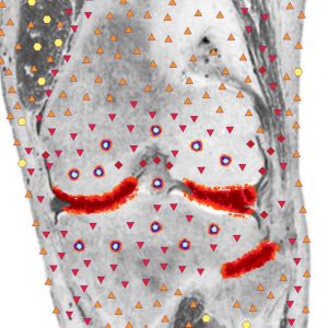
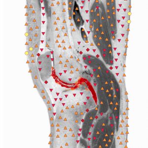
The NLS investigation was carried out using “Metatron” device equipped with a 4.9 GHz trigger sensor. Changes in the joint capsule were evaluated in virtual shots in sagittal planes above and below the kneecap and along the posterior surface of the joint. Frontal planes along the lateral surfaces of the joint were used to define the exact condition of menisci, articular cartilages and changes in the synovium.
It is traditionally believed that in the articular cartilage degenerative changes start off with a rupture of the articular matrix and degeneration of chondrosites. Therefore, during the NLS examination special attention was paid to changes in the articular cartilage. In the examinees of the test group, the articular cartilage looked like a hypochromous strip (1-2 points according to the Flandler’s scale). Two patients were found to have an articular cartilage of a heterogeneous chromogenic pattern, 3-5 points, in the initial phase of the disease with small hypochromogenic nidi (1-2 points) present. No radiological changes in the joints were detected for this group of patients.
In 14 (28.0%) patients in the second clinical phase of the disease, the chromostructure of the cartilage was heterogeneous and some moderately hyperchromogenic structures (4-5 points) were detected as well as hypochromogenic inclusions (1-3 points) of a small diameter. In 21 (42%) examinees in the third phase of the disease, the hyaline cartilage looked as a hyperchromogenic strip (5-6 points).
In 10 (20.0%) patients in a third clinical phase of the disease the articular cartilage was visualized as a distinctly hyperchromogenic linear structure (6 points) with vertical fissures present (4-5 points).
Depending on the phase and duration of the disease, a spectral similarity (D 0.189 to 0.621) could be visualized to the etalon process “osteoarthritis deformans”.
The X-ray pictures detected a moderate constriction and deformity of the joint space as a primary sign of the articular cartilage distraction in 22 patients and considerable constriction in 12 patients. Subcartilaginous osteophyte was very important for osteoarthritis patagenesis. Formation of subcartilaginous and epiphyseal cysts started already in the initial phases of the disease (71.0% of the patients). According to NLS-investigation, the cysts were located subcartilaginously in the lateral regions of the bone, 1.0-3.0 mm deep and were as many as 4 to 12-15. Standard X-ray pictures of knee joints displayed some changes in the subcartilaginous regions of the bone, like cysts and fibrosis, only in the second phase of the disease.
A very important role in the osteoarthrosis deformans development was attributed to the condition of the synovium and articular capsule. With the progress of the disease and changes in its phases, a cartilaginous detritus with antigenicity was formed on the articular surfaces. That often led to the inflammation of the synovium and its fibrosis. As a result, the synovium produced an inadequate fluid, which in turn impaired the cartilage nutrition with its ensuing degeneration.
The synovium in healthy persons (test group) was visualized as a hypochromogenic linear structure (1-2 points). The first and second phases of the disease saw a steady rise in its chromogenic pattern in 14 (28%) patients (3- 4 points). In 32 (62%) patients in the third phase of the disease the chromogenic density of the membrane reached 4-5 points throughout the phase with at most 3 or 6 hypochromogenic inclusions. In three patients with an aggravated form of the osteoarthrosis deformans (the forth clinical phase) the synovium looked like a distinctly hyperchromogenic structure (4 points) with areas of a reduced entropic density (3-4 points).
Changes in the membrane structure were always concomitant with synovitis with a limited amount (mostly in the upper enstrophe in 28.0% of the patients) or a great amount (in all regions of the joint – in 68% of the patients) of fluid free of sediment and additional inclusions.
Depending on the phase and extent of pathological changes in the joint affected by osteoarthrosis a change in the joint capsule structure also took place. Only in the first phase of the disease did the joint capsule structure remain normal. In the second phase of the disease, especially with synovitis in evidence, the chromostructure was assessed at 4-5 points in 14 (28%) patients and in the third and forth phases of the disease – up to 6 points in 34 (68%) patients.
Roentgenographic evidences of synovitis and changes in the paraarticular soft tissues were detected in some patients only in the third and fourth phases of the disease.
Thus, the analysis showed that the NLS-investigation had an advantage over conventional roentgenologic methods in terms of early detection of degenerative changes in the articular cartilage.
Overall, the NLS-method sensitivity in the early phase of the disease amounted to 82%, specificity to 85% and accuracy to 86%. The sensitivity of standard radiography in two projections was 68%, specificity 54% and accuracy 78%.