Vladimir Nesterov
Full member of Academy of Medical and Technical Sciences, Academician,
President of International Academy of non-linear diagnostic systems.
Among different kinds of lung disorders, special attention has been paid over the last years to diffuse infiltrative lung diseases (DILD), which is largely accounted for by some problems in their timely diagnostics and treatment.
Most diffuse lung diseases involve in the pathological process both the interstitial tissue and the respiratory tract and alveola. In this connection this type of pathological processes should be defined rather as diffuse infiltrative than as interstitial diseases. Despite of the polymorphism of clinicomorphological manifestations of DILD, most of them start with productive alveolitis (in contrast to the exudative alveolitis in the case of a pneumonia) with fairly stereotyped changes in the lung interstice in the form of inflammatory infiltration with different degrees of intensity. Subsequently a fibrosis develops that can have different rates of progression. A “cellular lung” pattern is the final phase of the development. It should be noted, that some infectious diseases of certain etiology (like tuberculosis, histoplasmosis, etc.) and particular malignant tumors (lymphogenous carcinomatosis, bronchioloalveolar cancer) do not directly belong to interstitial lung diseases but are similar to them in terms of manifestation.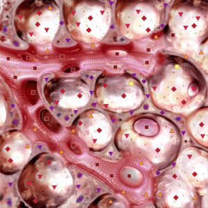
The clinical evaluation of patients with a suspected DILD is a complex problem. Nonspecific symptoms and in some cases signs detected during chest examination may be characteristic of a multitude of acute or chronic lung diseases that involve the interstitial tissue, respiratory tract or alveola. An extremely heterogeneous group of diseases represents DILD. The DILDs have been described in over a hundred possible versions, however in clinical practice, only about 10 or 15 conditions are most common and it should be noted that sarcoidosis and various cases of lung fibrosis occur in clinical practice in 35-50% of all DILDs. Besides, acute diffuse lung processes in patients with reduced immunity (also in combination with HIV-infection) are likely to have a great number of infectious and non-infectious varieties, which X-ray evaluation is found to be difficult.
Unfortunately, the capabilities of conventional roentgenography for patients with a suspected DILD appear to be limited for the sensitivity and specificity of the method prove to be insufficient. The data on 458 patients with a histologically confirmed DILD were studied. The chest radiographs for 10% of the cases turned out to be normal. Among 86 patients affected by DILD no pathological change was detected in 50% of the patients with histologically proven bronchiectasia and in over 20% of the patients with emphysema shown on X-ray shots. Radiography may equally show false positive results of the investigation. We have discovered that in 10-20% of the patients with the x-ray-confirmed signs of DILD no changes were detected during the lung biopsy.
The computer nonlinear diagnostics (NLS) is one of the promising methods of diagnosing lung diseases of today.
NLS appreciably improves the communication of the fine morphological elements in the lung tissue and opens up new opportunities for recognizing interstitial diseases of the bronchoalveolar system.
NLS has a high sensitivity in detecting fine interstitial lesions of the parenchyma and small nodules.
The results of the investigations prove that NLS has a better sensitivity in detecting both acute and chronic diffuse lung diseases. The sensitivity of the NLS diagnosis in detecting lung diseases makes 85% as compared to 70% in chest radiography.
The accumulated experience too, gives additional grounds to assert that NLS is a highly efficient method for diagnosing a wide range of various diffuse lung diseases, DILD included, and excels the “classic” chest radiography by sensitivity.
It should be noted that the high sensitivity of the NLS-method is achieved without sacrificing the specificity and diagnostic accuracy of the method. In patients affected by DILD the NLS specificity amounted to 96% as opposed to 76% in radiography. In particular, the high sensitivity (87-92%) and specificity (88-94%) of NLS were demonstrated in bronchiectasia diagnostics.
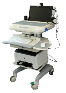
Although NLS is a more sensitive method as compared to chest radiography, its sensitivity in lung disease diagnostics is not absolute and the fact that no radiological changes were detected by NLS may lead to precluding lung disease in patients who actually suffer from DILD. 100 patients were examined by means of the NLS with “Metatron”-4025M device, 86 of them affected by DILD and 14 having no pathological change in the lungs. Despite a high value of NLS sensitivity and specificity, for 4% of the patients with biopsy-detected lung diseases the results were interpreted as being normal. On the other hand, the NLS was proven a high-accuracy technique for precluding acute lung diseases in patients with immunodeficiency.
Some examination data were studied for patients with a bone marrow transplant and clinical symptoms of fever of obscure genesis. The authors demonstrated high reliability of the NLS in determining fungal infection in 20 of 24 cases. Besides, the fact that no changes were detected during NLS lung examination allows to assume that the fever was caused by bacterial or fungal infection of extra pulmonary genesis.
It is also a proven fact that the sensitivity with NLS is higher than with standard computer tomography. We examined 150 patients. Using conventional CT (10 mm collimation) and NLS, we found that NLS had a higher sensitivity in recognizing pathological changes in the lung tissue.
Due to its high sensitivity, NLS should be used to define lung diseases in patients with a normal or obscure aspect of disease who have a pulmonary disturbance or symptoms that suggest an acute or chronic diffuse lung disease.
Even with certain clinical signs in evidence, the diagnostic accuracy of classic radiography in patients affected by DILD appears to be limited. The reason is both superposition of the image in the radiograph and low contrast of minute lung structures. NLS is free of these aspects, which is why it is reputed to be a more efficient method for recognizing diffuse lesions of lung tissue as compared to both radiographic survey and conventional computer tomography.
Besides, having a higher sensitivity, specificity and diagnostic accuracy, the NLS method can become a determining factor in evaluating the activity of a pathological process in patients affected by DILD. In certain cases NLS can be used not only to define the presence or absence of a pathological process or the extent to which it has spread, but also to collect information about the reversibility of changes (in an acute or active phase) as compared to irreversible (fibrotic) changes in the lung tissue. Moreover, since NLS can accurately identify the imponderable activity of a pathological process in the lungs, it can be employed to evaluate the efficiently of the treatment given to the patients.
The conventional methods for evaluating disease activity, such as transbronchial lung biopsy (TBLB), bronchoalveolar lavage (BAL), chest radiography, gallium lung scanning and functional lung tests are insufficiently reliable in evaluating the activity and in terms of prognostication. So the open lung biopsy (OLB) is still the choice method for both diagnosing and evaluating the process activity. We were able to prove, that signs detected in patients by means of NLS can provide some valuable information and be significantly important in defining the activity of a pathological process.
In terms of its prognostic value NLS is now advancing to the foreground leaving behind some functional lung tests, BAL and even OLB, because it allows to assess a lesion of actually the whole lung parenchyma as compared to a separate biopsy sample. Moreover, NLS can become an accurate noninvasive method for evaluating the efficiency of the administered treatment.
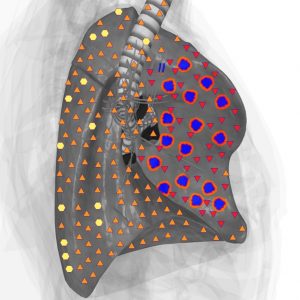
Sarcoidosis is one of the most common interstitial lung diseases of unknown etiology. In typical cases, granulomas are formed in fine lymph vessels or beside them, afterwards the granulomas self-organize that causes lung tissue fibrosis. A number of researchers considered the NLS potentials in defining the process activity in patients affected by sarcoidosis. The main activity indicator is the presence of small nodules and to a lesser degree their distribution and occurence in the lung tissue. Unfortunately, despite the difference between reversible and irreversible changes detected by NLS for patients having sarcoidosis, the potentials of NLS in assessing the process activity have not been studied well enough.
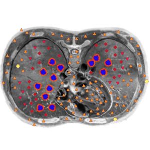
Among different indications in favor of NLS application, the use of this method in lung biopsy is probably the most important one. Biopsy is a very essential diagnostics technique, which allows defining the nosology of a lung disease, its activity level and phase. The diagnostic value of biopsy to a certain degree depends on its method and the type of DILD. The authors proved that TBLB was diagnostically informative for only 20 patients of 53 (38%) who had DILD in evidence; in 33 such patients (62%) TBLB displayed normal lung tissue or nonspecific changes. At the same time, OLB made a specific diagnosis of DILD in 92% of cases. In DILD-affected patients, TBLB proved to be most informative for patients having sarcoidosis or lymphogenous carcinomatosis, because these lesions have largely peribronchial tissue involved and are therefore most accessible to TBLB. Diagnostically OLB appears to be more accurate, but it also has certain complexities because lung tissue is sampled from a small sector of the lung, which might not reflect the changes occurring in the rest of the lung tissue. Many diffuse diseases affect lung tissue irregularly so the pathologically altered parts of lung parenchyma may be located among normal lung tissue. Besides, the same lung may contain both active manifestations of the disease and fibrotic changes of long standing. For an accurate diagnosis and assessment of the clinical progress of the disease, the right choice of a biopsy sample is very important. During biopsy, NLS helps to collect accurate data, indicating active areas of a pathological process. By using NLS, the areas affected by lung fibrosis in its final phase, with “honeycomb lung” formed, could be skipped during biopsy sampling. In addition, NLS may prove to be vitally important for choosing the most effective technique (TBLB, BAL or OLB) for making a histological diagnosis.
Radiography remains the most accessible method for diagnosing DILD yet its informational content appears to be not sufficient. Making a correct diagnosis necessitates a combination of laboratory, functional and radiological investigations as well as some invasive methods, each of them having its own substantial limitations.
NLS-diagnostics is a method that greatly improves identification of diffuse infiltrative lung diseases and as such, it should become an integral part of an integrated investigation.