Vladimir Nesterov
Full member of Academy of Medical and Technical Sciences, Academician,
President of International Academy of non-linear diagnostic systems
Screening NLS-investigation detected two cases of lung abscess in feverish patients who were complaining of pain in the right hypochondriac region. The patients were subjected to echography in order to exclude an abdominal cavity pathology.
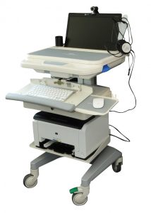
The NLS examination was conducted by means of “Metatron”-4025M device equipped with a digital trigger sensor of 4.9 GHz.
Patient N., aged 57, was admitted to the therapeutic department. He was complaining of a weeklong fever with a temperature of up to 40.0°C, a moderate non-productive cough and pain in the right hypochondriac region because of catching a cold. He came to see a doctor ten days after falling ill. The anamnesis showed a bilateral pneumonia 14 years earlier. The clinical blood analysis indicated an increased leukocyte content – up to 18.7 x 10⁹ with a leukogram left shift. The common urinalysis showed no deviations.
Physical examination: vesicular pulmonary respiration, weakened in the lower sections on the right with no rhonchi. Tongue dry, white furred. Belly soft, with frank painfulness in the right hypochondriac region. No symptoms of peritoneum irritation in evidence. Pasternatski symptom negative on the right and left.
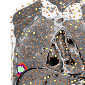
The NLS-investigation of the abdominal cavity did not detect any signs of pathology in the liver, gall bladder or pancreas. On the right there were visualized high chromogenety areas in the diaphragmatic pleura (4-5 points according to Fleindler’s scale) and an image of voluminous formation in the right lung was acquired (5-6 points). On the dorsal thoracic wall, there was an image of an enhanced chromogenic formation (6 points) of a heterogeneous internal structure, sized 80x65x54 mm with achomogenic central area (0 points).
The lung tissue around the nidus had a higher chromogenic density (4-5 points) because of infiltration. A spectral similarity to the “lung abscess” etalon (D=0.312) was detected. The investigation of the left lung and pleural cavities did not detect any structural changes. Conclusion: NLS signs of developing abscess in the right lung.
Control radiological investigation arrived at the conclusion: an abscess in the lower lobe of the right lung in progress.
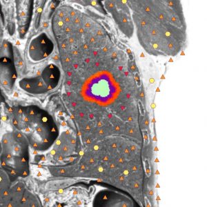
A repeated NLS examination was conducted 10 days later. It visualized a rounded hyperchromogenic formation with uneven outlines with achromogenic zones inside, sized 81x60x51 mm. The chromogenic density of the lung tissue around the nidus was somewhat higher (due to infiltration), and the folia of the visceral and parietal pleuras were blackened in the lower sections of the right lung.
The patient was offered a further therapy in a specialized surgical department, which he turned down. 3 weeks later, after some anti-inflammatory therapy, control NLS examination was performed. During the examination the patient complained of coughing with a profuse sputum discharge. His body temperature was normal, the clinical blood analysis indicated a leukocyte count of 8.6×10⁹, the differential blood count was within the standard, and ESR grew up to 37 mm/h. The
NLS-investigation visualized a rounded formation with even outlines, increased chromogenic density and achromogenic internal structure sized 47×43 mm. The chromogenic density of the lung tissue around the perimeter decreased (because of reduced infiltration).
At the patient’s urgent appeal he was discharged from hospital for further outpatient treatment. Later he underwent two check examinations.
Patient M., aged 63, was examined by means of the NLS-method in order to exclude liver or gall bladder pathologies.
NLS-investigation of the lung and pleural cavities was carried out. In the left lung and pleural cavities it found no signs of pathology in evidence. In the right lung in the IX, X and XI hypochondria (from the paravertebral line to the scapular one) it parietally visualized a formation having an increased chromogenic density and sized 85×60 mm with uneven outlines and heterogeneous structure (due to inclusions of a decreased chromogenic density, sized 3-4 mm). The chromogenic density of the lung tissue was not increased. Conclusion: NLS signs of an abscess in the right lung?
Radiological conclusion: abscess in the lower lobe of the right lung.
The patient had check NLS-investigations conducted against the background of anti-inflammatory therapy.
With the NLS-investigation performed 10 days later the formation looked rounded, had even outlines, an increased chromogenic density (4-5 points) and a achromogenic internal structure. Around the perimeter of the nidus the lung tissue had an increased chromogenic density (3-4 points), because of infiltration. The formation measured 73x50x60 mm.
The NLS-investigation 2 weeks later did not detect any positive dynamics from the administered anti-inflammatory therapy. The patient was sent to a specialized department for operative treatment.
The presented clinical observations once again confirm that the NLS-investigation at lung diseases is not used in clinical practice as often, as it deserves. Besides, the dynamic NLS-observation of patients affected by lung diseases allows assessing the efficiency of the employed therapy and reducing the radiation load both on patients and on the medical personnel.