T. Van de Kerkhof, М. Bawerman, C. Murphy
UCT Private Academic Hospital, Capetown
Adrenals tumors are registered comparatively rarely, but it is know that they are most frequently hormone-active and even at small sizes lead to various endocrine disorders. However sometimes in clinical practice there are adrenals tumors without clinical symptoms and vague complaints of patients.
References and our own experience show that period from appearance of the first symptoms till diagnosing varies from 6 months to 2 years.Early diagnostics of adrenals tumor has become an important clinical issue, which may be solved by application of non-linear diagnostics (NLS) along with CT, MRI and angiography.
Today NLS-graphy is one of the most informative and prospective methods of diagnostics. Thanks to easiness of application, high informativity and safety NLS-graphy becoming a method of choice even at the first stage of hardware methods of diagnostics. NLS method of study allowed to increase significantly percentage of early and accurate diagnostics of adrenals tumors. Along with introduction of the latest devices of a fourth generation it became possible to register any space-occupying formation sized about 0.2 cm, which exceeds CT in accuracy of diagnostics.
The objective of our work is to study possibilities of NLS-graphy in diagnostics of adrenals tumors and to confirm that with proper methodological approach and availability of modern NLS-graphy equipment it is possible to diagnose space-occupying formation at least as good as with CT and MRI.
Material and methods
Clinical data: from June 2012 till May 2013 we studied 23 patients aged from 25 to 64, in which, in accordance with complaints analysis and clinical-laboratory data, we assumed adrenals tumors. All patients were subjected to kidneys and adrenals study in accordance with standard method with use of Metatron-4025 device (IPP, Omsk) with 4.9 GHz generator frequency and professional software Metapathia GR Clinical allowing to apply three-dimensional visualization of adrenals. At the same time we carried out spectral-entropic analysis (SEA) of adrenals tissue structure, which helped to identify pathomorphological picture of adrenals affection by comparison of a pathology spectrum to etalon processes.
Analysis of our findings evidences that correct diagnosis was set in 22 cases of 23 (95.6%). In one patient with Itsenko-Cushing syndrome NLS-graphy detected small hyperchromogenic formation (30-35 mm) in superior pole of right kidney, which had homogeneous internal structure and was identified as benign tumor of adrenal gland. CT was not applied, because we saw a typical clinical picture of Itsenko-Cushing syndrome, confirmed by SEA. During surgery we found nidal macronodular hyperplasia of a gland. SEA results were confirmed by histological study of removed formation.
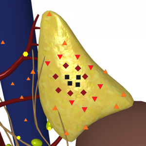
Pic. 1. 3D NLS-graphy. Space-occupying formation, less than 1.5 cm in diameter in right adrenal gland.
According to references there are diffuse and nidal types of hyperplasia of adrenals. Nidal hyperplasia is divided into micro- and macronodular. NLS-sings do not allow to differ nidal hyperplasia from adrenals tumor, so in this case only application of SEA allows to avoid diagnostic mistake.
In 17 of 23 cases we applied CT and acquired data coincided with NLS data within the accuracy of 3-5 mm. 9 of 23 patients were subjected to surgical removing of adrenals tumors and in all cases NLS and SEA data were confirmed by operative data and histological research.
Size of revealed tumors varied from 2 to 10 cm in diameter. Lesser tumors had homogeneous isochromogenic structure, in 4 cases, when tumor was larger than 4 cm, internal structure was represented by erratic alternation of high and low chromogeneity areas due to necrosis zones, degenerative changes and calcification, which was confirmed during histological researches of removed tumors.
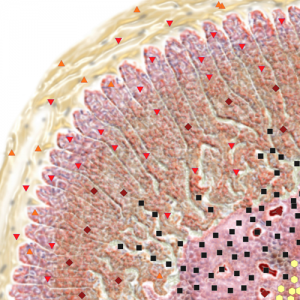
Pic. 2. NLS-ultramicroscanning. Adrenal tumor with degeneration in a center.
According to our data differential diagnostics of benign and malignant tumors of adrenals is extremely difficult. Only in case of heterogeneous internal structure of a formation, limited motility at forced breathing, presence of enlarged regional lymph nodes, metastases into other organs, we may assume malignant character of detected formation. Results of SEA in this case are diagnostically important aspect.
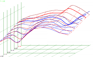
Pic. 3. Spectral-entropic analysis. Corticosteroma.
Therefore diagnostics of adrenals tumors is quite difficult, i.e. their clinical manifestations are diverse. It hard to overestimate possibilities of NLS diagnostics with SEA, which thanks to easiness of study, safety and high informativity allowed us to set a diagnosis quickly and accurately in 98% of cases. The study has shown that NLS-method may become equal with CT in studies of adrenals tumors. Patients, who have typical for adrenals tumors complaints, first of all must be subjected to NLS-graphy. At detection of NLS signs of adrenals tumors, these patients must be subjected to SEA and in case of positive results – further detailed study with CT and MRI, this scheme will shorten patients check-up time significantly and allow to carry out proper and well timed treatment.
References
1. Grigoryan S.V., Kuchinsky G.А., Molchanov G.P. Peculiarities of clinical course and treatment of gigantic tumors of adrenal cortex. Archives 1989, v.61, No.1, page 124. 1988.
2. Yeh H.C. NLS-graphy of the adrenal glands: normal glands and small masse. A.J.R. 2013. t.85, p.87-97.
3. Gunter R.W., Muller-Leise. Radiologie of Adrenals.In Lang E.K. ed Radiologie of theupper urinary tract. Berlin,Heideberg,New York,Springer-Verlag. 1988.
4. Lukashenko B.I., Georgadze Т.P. Possibilities of NLS-scopy in diagnostics of adrenals tumors // Collection of scientific works of the Institute of Practical Psychophysics “Actual aspects of NLS-diagnostics”. Volume I. М.: Katalog, 2006, p. 60-64
5. Nesterov V.I. 3D NLS diagnostics. Prospects of development // Collection of scientific works of the Institute of Practical Psychophysics “NLS-technologies in medicine – prospects of development. Volume III. М.: Katalog, 2010, p. 5-8.
6. Nesterova V.I., Shaposhnikov L.V., Yankina L.А., Kozhemyakin О.R. Application of 3D NLS-diagnostics in oncology. New trends and prospects of development // Collection of scientific works of the Institute of Practical Psychophysics “NLS-technologies in medicine – prospects of development. Volume III. М.: Katalog, 2010, p. 9-12