U.N. Manuilov
Omsk regional oncologic dispensary
INTRODUCTION
Cervix carcinoma incidence rate growing is marked in the recent years both in Russian Federation and generally in the world. Increasing of cervix carcinoma incidence rate in women is a disturbing fact, because this group of patients is not only a significant part of reproductive female population, but socially active group also.
The main role in treatment of cervix carcinoma patients belongs to surgical intervention and ray therapy. Nowadays methods of combined and integrated treatment are developed and introduced into a practice.
Application of advance technologies for treatment requires support of a modern diagnostics. A group of hardware visualization is represented by the following methods: ultrasound research, computed tomography, magnetic-resonance imaging. Recently developed method of three-dimensional non-linear (NLS) diagnostics has several unquestionable advantages over other methods. 3D NLS-diagnostics method is safe for a patient and a doctor in regard to radiation exposure, has sufficient accuracy and high reliability of acquired results. 3D NLS-study of patients suffering from cervix carcinoma may be used both for diagnostics of an oncological process and for monitoring during treatment process in order to correct promptly or evaluate efficiency of treatment measures. Application of modern devices for NLS-diagnostics allows to evaluate condition and a structure of uterine cervix, dissemination of a process into uterine body, ovaries and urinary bladder. NLS-study of abdominal cavity and retroperitoneal space organs allows to evaluate dissemination of a process, detect presence of metastatic affection of liver, retroperitoneal lymph nodes.
Spectral-entropic analysis (SEA) of a tumor makes possible to understand pathogenetic mechanisms of tumor growth and to develop anti-tumor strategy. NLS-method makes possible to acquire multidimensional picture of a researched object and its vascular tree in real-time mode simultaneously.
Combined NLS-diagnostics of cervix carcinoma with application of ultramicroscanning with SEA allows to evaluate size and character of a tumor, which to a considerable extent defines treatment tactics at every stage of the treatment.
The objective of the study is to define possibilities of 3D NLS-research with ultramicroscanning and SEA in evaluation of patients with cervix carcinoma treatment efficiency.
MATERIAL AND METHODS
The study was carried out with participation of patients suffering from cervix carcinoma of 2nd and 3rd stage, treated in Omsk regional oncologic dispensary.
During the study we used “Metatron”-4025 systems with 4.9GHz high-frequency sensor and “Metapathia GR Clinical” professional software with features of three-dimensional visualization and evaluation of microscans.
At the first stage of the study all cervix carcinoma patients were subjected to survey transabdominal ultrasound check-up of small pelvis organs in V-mode after preliminary natural filling of an urinary bladder, invasion of urinary bladder was checked. At the second stage of the study we carried out three-dimensional NLS-study of small pelvis organs. Three-dimensional NLS-graphy helped to detect presence of uterine cervix damages and evaluate condition of endocervix. Lateral virtual cross-cut gave images of anterior and posterior walls of an uterus; frontal cross-cuts gave full picture of left and right sides of an uterus condition and condition of parametrial tissue. In three-dimensional NLS-angiography mode we studied spatial virtual picture of vascular structures of uterine cervix. Simultaneous 3D-visualization of the whole organ allowed to get clear idea of tumorous nidus volume. At the same time SEA was applied, it made possible to specify pathomorphological picture of uterine cervix affection by similarity of tumor’s spectrum and etalon processes. At the third stage of cervix carcinoma patients study we carried out NLS-study and SEA of liver and retroperitoneal space to detect remote metastases. NLS-studies complex was applied in accordance with the protocol developed be our team: before a treatment, during a treatment, after a treatment and in 6 months after a treatment.
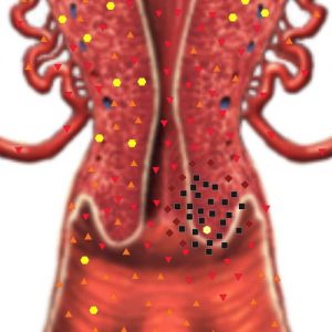
Pic.1 Two-dimensional NLS-graphy of uterus’ posterior wall. Cervix carcinoma.
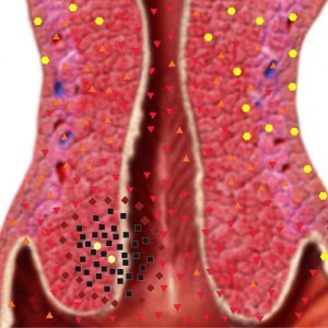
Pic.2. Two-dimensional NLS-graphy of uterus’ anterior wall. Cervix carcinoma.
RESULTS
Altogether 51 patients were studied. The following parameters were studied: age, morphological structure of a tumor, tumor growth form, variant of extra-organ spreading of a tumor. Age of all studied patients varied from 26 to 79 years (49.7 years old on the average). In all patients cervix carcinoma diagnosis was morphologically verified. In 92% of patients squamous cell cancer of various differentiation prevailed, in the rest of the patients adenocarcinoma was detected.
21.6% of patients had 2nd stage cancer, 78.4% – 3rd stage. Study of cervix carcinoma growth regularities showed: in 37% of patients combined form was detected, in 29.6% – endophytic form, in 31.4% – exophytic form and in 2% – infiltrative-ulcerous one. Variants of extra-organ spreading of a tumor: vaginal- parametric variant was detected in 68% of patients, vaginal- parametric-utricular – in 10% and parametric – in 22% of patients.
NLS-study of uterine cervix before a treatment found changes of uterine cervix structure: hypochromogenic nidi (of 5-6 points according to Fleindler’s scale) of various shape and size without strict contours and/or lesser hyperchromogenic inclusions of roundish shape. As a rule contours of uterine cervix are indistinct, uneven and tuberiferous. Average and high degrees of vascularisation were detected, high degree of vascularisation prevailed.
3D NLS-study of small pelvis organs spreading of tumorous process into uterus body was detected in 15.7% of patients, into ovaries – in 10%. NLS-ultramicroscanning and SEA of abdominal cavity and retroperitoneal space organs revealed affection of retroperitoneal lymph nodes in 4% of patients.
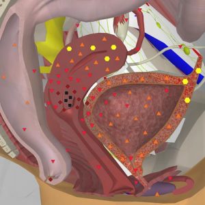
Pic.3 3D NLS-graphy. Cervix carcinoma.
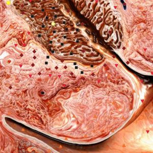
Pic.4. NLS-ultramicroscanning. Cervix carcinoma.
Patients were subjected to a treatment depending on a stage, morphological structure, dissemination of a process and in accordance with Standards of malignant tumors treatment. At the 2nd stage of a disease – combined split ray therapy, chemoradiation in a dynamic mode, at the 3rd stage – combined traditional ray therapy, chemoradiation. Thermotherapy was applied 15-20 minutes before endocavitary gamma therapy, 5-6 sessions. Session time – 1 hour, temperature 48–52°С. Thermotherapy was applied as a modifier of endocavitary gamma therapy. Ray therapy was administered to 15.6% of patients, chemoradiation – to 58.8%, chemoradiation with thermotherapy – to 25.6% of patients.
During and after the process of treatment we detected changes of contours and structure of uterine cervix. At a positive response to applied treatment contours of uterine cervix get distinct and even lines, hyperchromogenic nidi in uterine cervix are not visualized. At application of SEA similarity with “Cervix carcinoma” etalon process decreased from 0.387±0.084 till 0.876±0.148. At negative dynamics contours become more even, nidal changes of uterine cervix remain. At application of SEA similarity with “Cervix carcinoma” etalon process has not decreased reliably – 0.387±0.084 and 0.412±0.091 correspondingly.
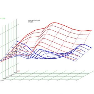
Pic.5. Spectral-entropic analysis. Cervix carcinoma. (D=0,227)
DISCUSSION
According to our studies cervix carcinoma is registered in women of socially active age. Morphologically squamous cell cancer of disseminated form prevailed. In general tumorous process was located in uterine cervix, excluding cases of spreading into body of uterus, ovaries and metastases into retroperitoneal lymph nodes.
In our study we included patients subjected to ray therapy, chemoradiation, chemoradiation with thermotherapy.
During and after the process of treatment we detected changes of contours and structure of uterine cervix.
We registered a tendency of uterine cervix volume decreasing in all patients.
It was the 3D NLS-study with SEA that allowed to evaluate efficiency of treatment in the most optimal way when applying various methods of treatment.
CONCLUSION
Complex of 3D NLS-study with SEA is a highly informative way of cervix carcinoma treatment efficiency monitoring.
REFERENCES:
1. Selected lectures on clinical oncology. Edited by RANS Academician V.I. Chissov and professor S.L. Daryalova. Moscow, 2000.
2. L.A. Kolomiyets, A.V. Vazhenin, M.I. Nechushkin. Focal cervix carcinoma: possibilities of radiotherapy // Modern oncology, 2005, Tome 7, No.4, p. 197-202.
3. I.B. Kaplanskaya, E.N. Glasko, G.A. Frank. Angiogenesis, intracellular contacts and stromal-parenchymal relations in normal and pathological conditions // Russian oncological journal. 2005, No.4, p.77-79.
4. Nesterova V.I., Shaposhnikov L.V., Yankina L.A., Kozhemyakin O.R. Application of NLS-diagnostics in oncology. New trends and prospects. // Collection of scientific papers of the Institute of Practical Psychophysics “NLS-diagnostics in medicine. Prospect of development”. Tome 3. Moscow. Katalog, 2010, p. 9-12
5. N.S. Karpov, I.V. Lemeshko. Comparative analysis of diagnostic value of NLS and MRI at invasive forms of cervix carcinoma // D computed NLS-graphy: Collection of works / Edited by V.I. Nesterov – Moscow, Publishing house Prospekt, 2012, p.43-47.